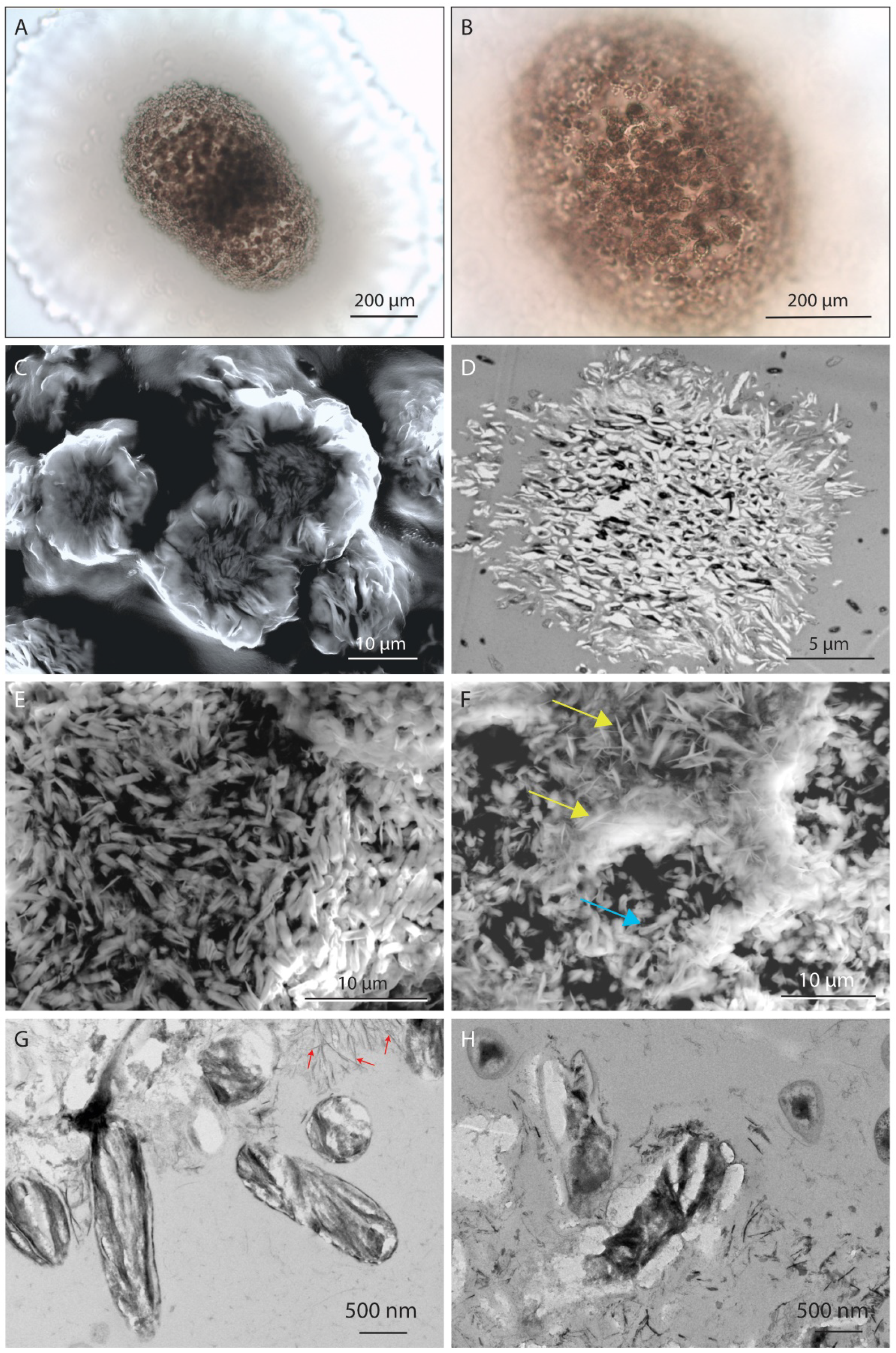
SEM (Scanning Electron Microscope) microphotographs of manganese

Download scientific diagram | SEM (Scanning Electron Microscope) microphotographs of manganese micronodules from the depth of 300 to 305 cm, size fraction 100-250 μm: а - micronodule with the frustules of Ethmodiscus, б - micronodule without admixture of valves of Ethmodiscus. from publication: Anomalies of rare elements in manganese micronodules from ethmodiscus oozes in the Brazil basin of the Atlantic Ocean | The composition of manganese micronodules from miopelagic clays and Ethmodiscus oozes of the central part of the Brazil Basin (station 1537, R/V Akademik Sergei Vavilov) is considered. Micronodules were recovered from >50 μm fraction of sediments from the depth intervals of | Manganese, Brazil and Atlantic Ocean | ResearchGate, the professional network for scientists.

Electron Microscopy Techniques, Strengths, Limitations and

Scanning electron microscope micrographs of the black deposit of
Scanning electron microscope (SEM) (a) and transmission electron
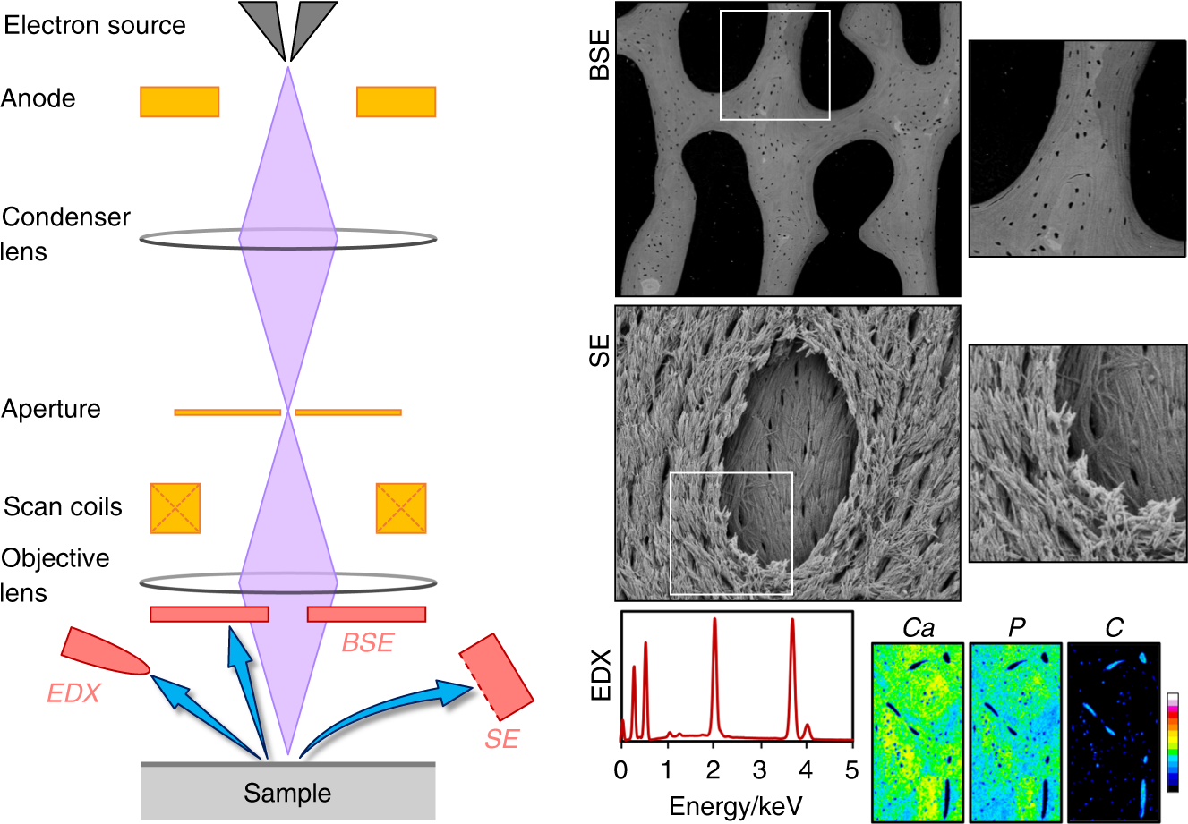
50 years of scanning electron microscopy of bone—a comprehensive
Scanning Electron Microscopy (SEM) - Surface Science Western

Representative scanning electron microscopy (SEM) images of (a
Power of Scanning Electron Microscopy and Energy Dispersive X-Ray
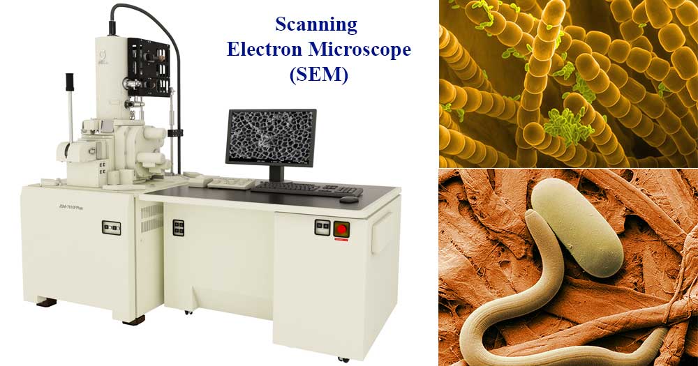
Scanning Electron Microscope (SEM)- Definition, Principle, Parts

Scanning Electron Microscopy images: (a) ZnO; (b) Mn(1 %):ZnO; (c
Minerals, Free Full-Text