Patient 3. Axial Spin Echo T1-weighted (A), Turbo Spin Echo T2-weighted

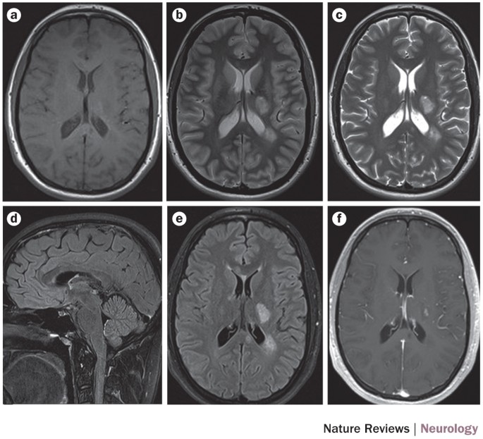
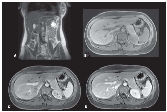
Download scientific diagram | Patient 3. Axial Spin Echo T1-weighted (A), Turbo Spin Echo T2-weighted (B), susceptibility weighted (C), diffusion weighted (b1000 and ADC map, D,E) and Turbo Field Echo T1-weighted after contrast administration (F) images. A left basal ganglia lesion also involving thalamus, corona radiata, deep temporal white matter and corpus callosum is appreciated. The lesion showed inhomogeneous signal intensity, with cystic and hemorrhagic components, and moderate enhancement. from publication: Germ cell tumors in male patients without gonadal involvement: Computed tomography/magnetic resonance imaging findings and diagnostic workflow | Germ Cell Tumors, Germ Cell and Embryonal Neoplasms and Gonads | ResearchGate, the professional network for scientists.

PDF) Single-shot, turbo spin-echo, diffusion-weighted imaging versus spin- echo-planar, diffusion-weighted imaging in the detection of acquired middle ear cholesteatoma

MRI artifacts still require significant care and attention
Basic and advanced mri imaging sequences in brain
MAGNIMS consensus guidelines on the use of MRI in multiple sclerosis—clinical implementation in the diagnostic process

Magnetic Resonance Imaging: The Underlying Principles
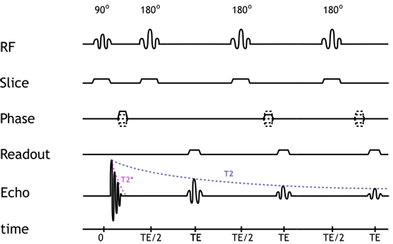
MRI Physics: MRI Pulse Sequences - XRayPhysics

Prostate Cancer: Comparison of 3D T2-Weighted With Conventional 2D T2- Weighted Imaging for Image Quality and Tumor Detection

Comparison of sagittal T2-weighted BLADE and fast spin-echo MRI of the female pelvis for motion artifact and lesion detection.
A comparison of a T1 weighted 3D gradient-echo sequence with three different parallel imaging reduction factors, breath hold and free breathing, using a 32 channel coil at 1.5T. A preliminary study

Detection of Sacroiliitis by Short-tau Inversion Recovery and T2-weighted Turbo Spin Echo Sequences: Results from the SIMACT Study

KJR :: Korean Journal of Radiology
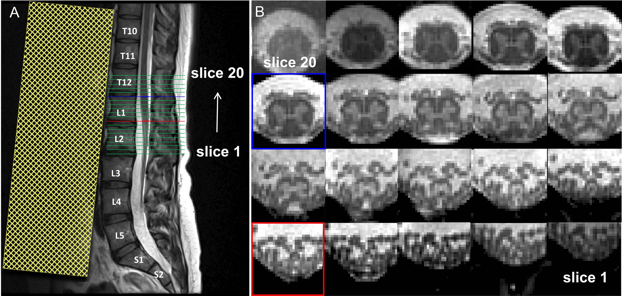
Optimized multi-echo gradient-echo magnetic resonance imaging for gray and white matter segmentation in the lumbosacral cord at 3 T
Radiologia Brasileira - RM de corpo inteiro: avaliação de protocolo em equipamento 3T com 48 canais em menos de 40 minutos. Resultados preliminares


)